
Eye diseases
Conjunctivitis
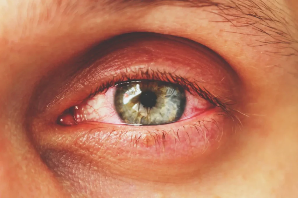
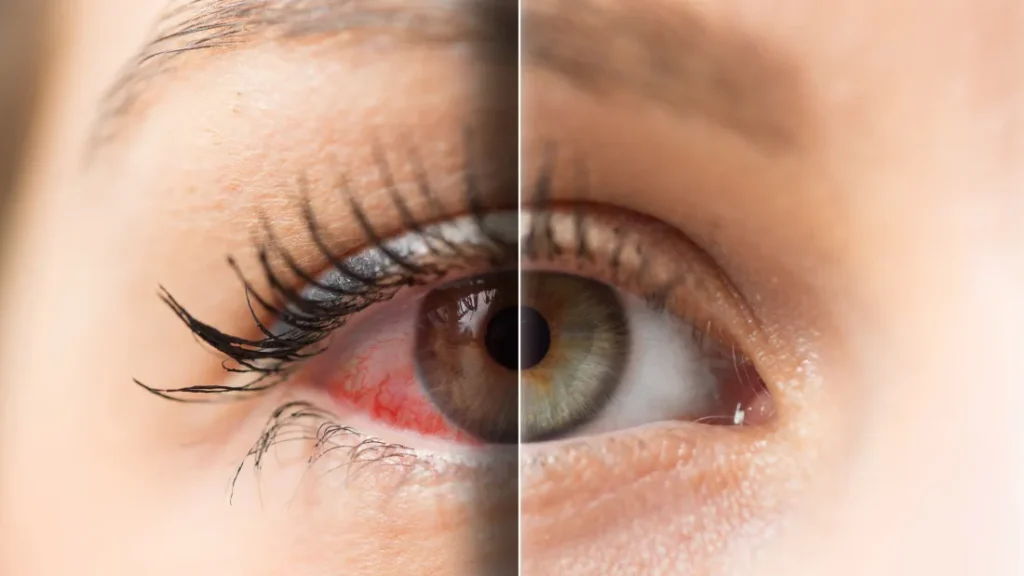
Conjunctivitis is an inflammation of the transparent membrane that covers the eyelid and the eyeball. This membrane is called the conjunctiva. The swelling and irritation of the conjunctival blood vessels makes them more visible and the sclera appear reddish or pink.
Conjunctivitis is most often caused by a viral infection. It can also be caused by a bacterial infection, an allergic reaction or – in babies – an incompletely opened tear duct.
Conjunctivitis
Symptoms
Although conjunctivitis can be irritating, it rarely affects vision, except in the case of inflammation spreading to the cornea when we talk about joint inflammation of the conjunctiva and cornea (keratoconjunctivitis).
Since conjunctivitis can be contagious, early diagnosis and taking certain precautions can help limit its spread.
Symptoms of conjunctivitis
Redness of the eyes
A burning and scratching sensation in the eyes
Increased lacrimation of both eyes
The appearance of secretions (watery or yellowish), which leads to "blindness" of the eyelids
Conjunctivitis
Treatment of conjunctivitis
In the case of viral conjunctivitis, there is no specific treatment.
The disease usually lasts 7-10 days and passes spontaneously.
In the case of a variant of viral conjunctivitis caused by certain types of adenovirus (epidemic conjunctivitis), the disturbances can last up to several weeks.
The therapy is carried out in the form of frequent washing of the eye with saline solution and the use of artificial tears in order to alleviate the symptoms.
Treatment of allergic conjunctivitis involves avoiding contact with the allergen, washing the eye more often, using cold compresses and artificial tears. If the disturbances are pronounced and persist, local and systemic antihistamines are used.
Bacterial conjunctivitis is treated with local antibiotics in the form of drops and ointments
Eye diseases
Cataract
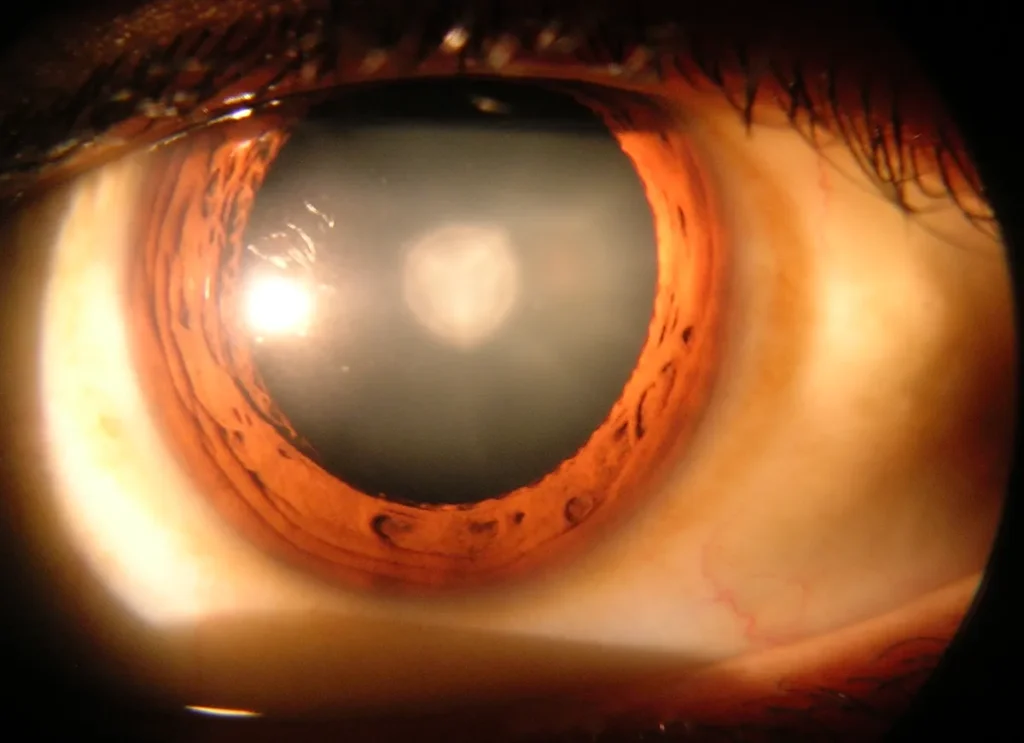
Cataract or cataract means any clouding in the lens of the eye which, when expressed, leads to a reduction in the quality of vision. Cataracts most often occur in people over 50 years old.The most common cause is aging, but risk factors include diabetes, smoking, exposure to UV radiation, and long-term use of corticosteroids.The patient usually does not notice the initial cloudiness until it affects the central parts of the lens or until the lens is completely clouded, when they can lead to a complete loss of vision.
Cataract
Types of cataracts
There are several types of cataracts, in relation to the cause that leads to the appearance of cataracts:
Senile cataract (cataracta senilis) or senile cataract is the most common form of cataract. Clouding of the eye lens is caused by the aging process of the body.
Congenital cataract (cataracta congenita) or congenital cataract. Babies are sometimes born with it or it develops in early childhood. Depending on the degree of clouding of the eye lens, this type of cataract sometimes does not interfere with the development of vision and then it is not necessary to operate on them. If a congenital cataract affects the development of vision, it is necessary to perform cataract surgery as soon as possible in order to prevent the occurrence of low vision (amblyopia).
Cataracts can also occur as a result of some other diseases. Diabetes is usually the cause, but this type of cataract can also occur as a result of long-term use of certain medications (most often corticosteroids).
Iatrogenic cataract (cataracta iatrogenes) caused by previous operations on other parts of the eye, radiation, drugs.
Traumatic cataract (cataracta traumatica) occurs as a result of eye injury. It can occur immediately after the injury or years later.
Complicated cataract (cataracta complicata) formed after other severe eye diseases or some other diseases of the body.
Cataract
Treatment of cataract
The only way to treat cataracts is through surgery. The clouded lens is replaced by an artificial, clear lens (intraocular lens).
Given that cataracts may not be the only factor in the eye that reduces visual acuity, and that a patient may have a diagnosis of glaucoma or macular damage in addition to cataracts, surgery may therefore provide a person with better vision, but still not full visual acuity.
This is a very quick, painless operation, which immediately eliminates the symptoms you have due to cataracts. There are many different types of intraocular lenses available today, and choosing the right intraocular lens is crucial.
Eye diseases
Glaucoma
Glaucoma is a group of eye diseases that lead to damage to the optic nerve, characteristic drops in the field of vision and gradual, progressive loss of vision.
The main risk factor for glaucoma is elevated eye pressure, but it is important to note that damage can develop even when the eye pressure is within normal limits, while on the other hand, elevated pressure can be present for years without damage occurring.
Glaucoma
Who are the risk groups?
Persons aged over 50 years
Relatives of glaucoma patients
Patients with diabetes and cardiovascular diseases
People with a previous eye injury that caused damage to certain structures in the eye.
People who have used corticosteroid therapy for a long time.
Glaucoma
Types of glaucoma
Many forms of glaucoma have no warning signs. The loss of vision is so gradual that you may not notice the change until the condition reaches a later stage.
Open-angle glaucoma - the most common type with a gradual increase in eye pressure. It progresses slowly, without specific symptoms.
Angle-closure glaucoma - angular, acute glaucoma. Symptoms are a sudden jump in eye pressure. It is caused by the blocking of drainage channels that release aqueous humor from the eye. Symptoms are severe pain in the eye, the appearance of rainbow colors around the bulb, blurred vision, severe headache followed by nausea and vomiting. An urgent examination by an ophthalmologist is required.
Glaucoma without elevated intraocular pressure – this type of glaucoma is characterized by nerve damage without elevated intraocular pressure.
Secondary glaucoma - a consequence of another eye disease, injury, systemic disease, tumor, use of drugs (corticosteroids).
Congenital glaucoma - occurs in the first months of life due to disturbances in the formation of eye structures for swelling of the aqueous humor. It is manifested by enlarged eyes and it is noticed that the child is bothered by light.
Glaucoma is a chronic disease that cannot be cured, but can be controlled by applying appropriate therapy. Glaucoma treatment depends on the type of glaucoma and the degree of damage to the optic nerve. Medicinal therapy (drops, tablets...), laser treatment and surgery are used.That is why it is extremely important to regularly perform eye examinations that include measurement of eye pressure. If glaucoma is detected early, vision loss can be slowed or prevented. If you have glaucoma, you will need treatment or monitoring for the rest of your life.
Eye diseases
Macular degeneration
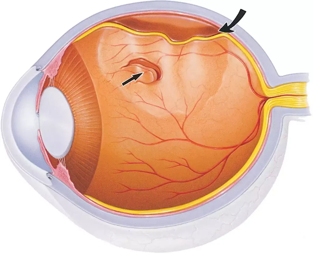
The yellow spot or macula is the part of the retina that is responsible for central vision. Macular degeneration is damage to the macula that occurs in old age, most often as a consequence of overall aging of the organism. The true cause of the disease is unknown, but it has been established that genetics plays a significant role. Familial occurrence of the disease is also known. With the increase in life expectancy, this disease is becoming a growing problem in ophthalmology.
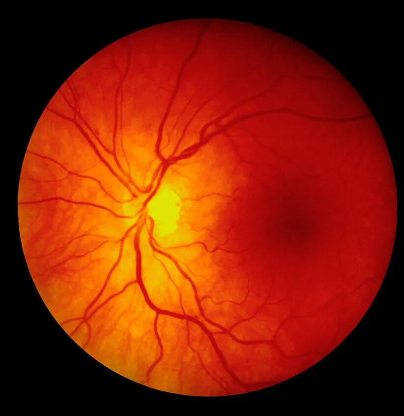
With this disease, the central part of the fundus (macula) is affected, and therefore this affects the central vision, which we need the most when we want to read something or focus on a certain object. Movement and navigation in space are preserved. Macular degeneration is not painful and the person very often does not notice any changes until significant damage to central vision occurs. The main symptom is blurred, blurred central vision that cannot be corrected with glasses.
Types of macular degeneration
We distinguish dry and wet forms of degeneration
The dry form of macular degeneration progresses slowly. Often, patients do not notice disturbances until the disease affects both eyes and the changes themselves are not pronounced enough to significantly impair reading. Regular check-ups are necessary, because this type of degeneration can turn into a wet (more serious) form of the disease.
The wet form of degeneration accounts for about 10% of cases of macular degeneration. Damage to central vision in this case is much more pronounced.
What distinguishes this form from the dry form of degeneration is the formation of pathological blood vessels, which are quite fragile, break easily, and as a result, liquid spills and bleeds in the central part of the fundus. Later, spontaneous scarring occurs when the decline in central vision is usually very pronounced (sometimes even final) and then we enter a phase in which there are not many therapeutic options.
Treatment of macular degeneration
Vitamin supplements and lifestyle changes
The use of appropriate vitamin preparations, prescribed by an ophthalmologist, slows down the progression of the dry form of the disease. It is also important to change the lifestyle in terms of a healthy diet with the use of large amounts of green fruits and vegetables, physical activity and smoking cessation.
Macular degeneration
Anti-VEGF terapija
Anti-VEGF injections represent a group of drugs that reduce the formation of newly formed fragile blood vessels. These injections are given in the eye, with the previous application of local anesthesia.
In our Center, we offer the possibility of using all modern preparations that are used in the treatment of this form of the disease.
Eye diseases
Retinal ablation
Ablation is a condition that occurs when the retina detaches ie. move from its normal position. Retinal detachment is an emergency because the cells are left without a continuous supply of oxygen and food through the blood, which results in their deterioration and carries the risk of permanent vision loss. That is why it is of crucial importance to report immediately for an examination and carry out treatment as soon as possible.
Symptoms of retinal detachment may include the following:
-reduced vision
- the sudden appearance of dark floating shapes and flashes of light in your vision
- loss of side vision.
Retinal ablation is treated surgically.
Ophthalomology Center Dr Raonić
In Ophthalmology Center - only the best.
We are proud to point out that in Ophthalomogy Center Dr Raonic operate specialists from the entire region. Our patients can also schedule appointments with dr Zoran Žikić - a specialist with decades of experience.