
Diagnostics
Optical Coherence Tomography (OCT)
Optical coherence tomography (OCT) is a modern, non-invasive diagnostic method that uses light waves to obtain detailed cross-sections of the retina and other eye structures.
Optical Coherence Tomography (OCT)
What is OCT?
Optical coherence tomography enables a three-dimensional display of the layers of the retina, which helps in accurate diagnosis and monitoring of eye diseases such as glaucoma, diabetic retinopathy and macular degeneration. This technology provides high image resolution and detailed information about the condition of eye tissues.
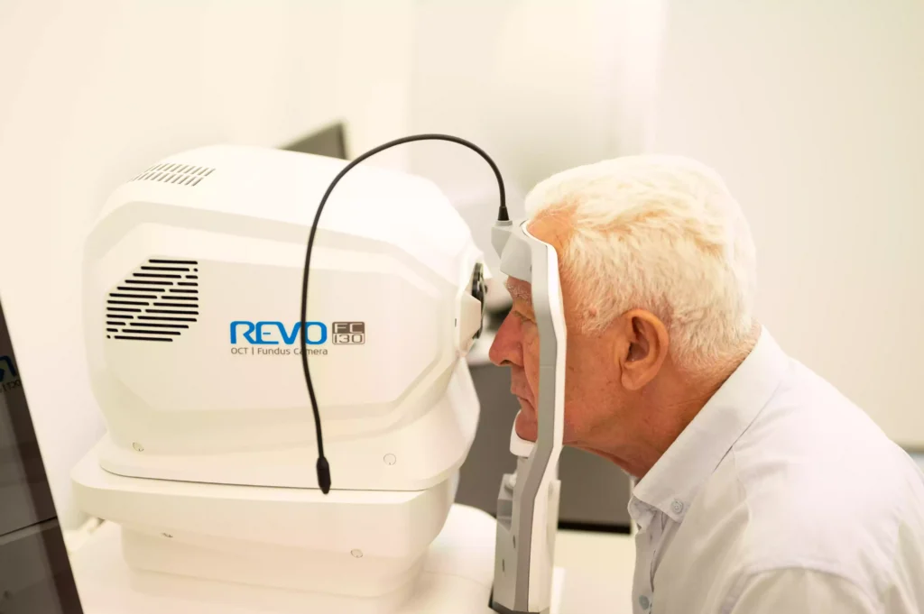
Our OCT device (REVO FC 130) offers multiple functions in one device, combining the potential of several devices. With just one OCT device, corneal-to-retinal changes can be measured, quantified, calculated and monitored over time, with the addition of a state-of-the-art color fundus camera (for anterior and posterior eye segment photographs) for a new level of diagnostic certainty.
Ophthalmology Center Dr Raonić
The capabilities of our OCT technology
- OCT angiography
- Precise measurement of corneal thickness (Pachymetry)
- Seeing a map of the cornea and existing corneal abnormalities, such as keratoconus (topography)
- Precise measurements of all structures inside the eye and the length of the eye (biometrics)
- OCT biometric module for monitoring myopia progression
- Determination of parameters of the lens to be implanted during cataract surgery (IOL calculation)
- Recordings/photographs of the fundus (fundus camera) without the necessity of dilating the pupil in patients in whom it is not possible (not indicated) or in situations where it is not necessary
- Imaging/photograph of the anterior segment of the eye